Anatomy Moment: The Suboccipital Muscles
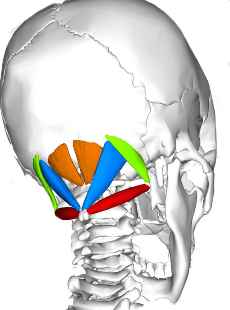
The suboccipital muscles are a group of eight tiny muscles - four on the right, four on the left - that attach the skull to the first and second vertebrae in the neck. Anatomical terms can give us a host of information about muscle shape or location, and, in some cases, function. Let’s explore what the suboccipital muscle names tells us:
Sub in anatomy lingo means under, and these muscles are under the occipital bone, or occiput, in the skull.
The occiput is essentially the back and base of the skull, and covers the corresponding occipital lobe in our brain.
The occipital lobe is responsible for visual perception, including colour, form, and motion. Correspondingly, the occipital muscles help make the tiny movements which position our head, and thus have a lot to do with our vision and how we take in the world as well.
TRY THIS:
Run your fingertips up the back of your neck until you feel the ridge or protrusion of your occipital bone - think where the back of the neck starts to become the skull. Gently press your fingers into the muscle right at that connection point. Close your eyes and move your eyes right and left behind your closed lids. Do you feel a “plumping up” under your fingers as the suboccipital muscles engage? These muscles don’t move our eyes, they position our head, but because head position has a lot to do with how we look at and see things, the suboccipital neurologically linked to eye position and will respond as you move your eyes!
Interestingly, the space where the suboccipital muscles live can get compressed or shortened by forward head posture, which we discussed in our previous blog on the longus colli muscle in the front of the neck. Long periods of looking at a computer screen (or textbook… or novel… but TBH computers are the big culprit for most of us!) often cause a forward lean to the head, which lengthens the longus colli and shortens the suboccipital space.
This shortened position and corresponding tension in the suboccipitals and other muscles at the back of the neck can be responsible for tension headaches and, in some cases, more intense headaches that feel like they’re almost behind the eye. Here’s a simple yet powerful release we love for these tension headaches:
TRY THIS:
Grab a yoga block (or thick book) and two pinky balls in a sock.
Place the pinky balls right where the neck and skull meet, on either side of the spine.
Let the weight of the head rest back, and try a few small yes nods.
Feel free to move the balls up or down a little bit - you may find an even juicier spot just a quarter centimeter away from where you initially put the balls.
Keep in mind: Never place the pinky balls directly on your spine. Make sure your neck feels comfortable - any strong sensation should be where the balls are providing pressure, not somewhere else in your neck.
Our necks are complex creatures, and understanding how the back, fronts, and sides are affected by our everyday activities as well as our exercises routines can be super helpful in finding long-term solutions to neck tightness and pain. We hope you’ve enjoyed our multi-part dive into the neck! For more neck care tips, come to class and let your instructor know how your neck is feeling today!
Let’s Move!
Book a group class or private session to use what you’ve learned: